Learning Outline
Skeletal System
The specific structures of the gross anatomy of the skeleton, such as names of specific bones and bone features are not covered in this outline.
If you want an optional overview of the bones of the skeleton click here.
Functions of the skeletal system
Support
Framework of body, holding other organs in place
Movement
Attachment sites for skeletal muscles
Movable joints; leverage for movement
Protection
Hard covering of thoracic organs, brain, spinal cord, other soft structures
Protection as in bodily defense against injury
Mineral and fat storage
Calcium & phosphorus salts stored in bone tissue
Yellow fat (yellow bone marrow) stored in bone cavities
Blood cell production
Hematopoiesis (hemato = “blood” poiesis = “making”)
Red bone marrow is blood-forming tissue inside some bones

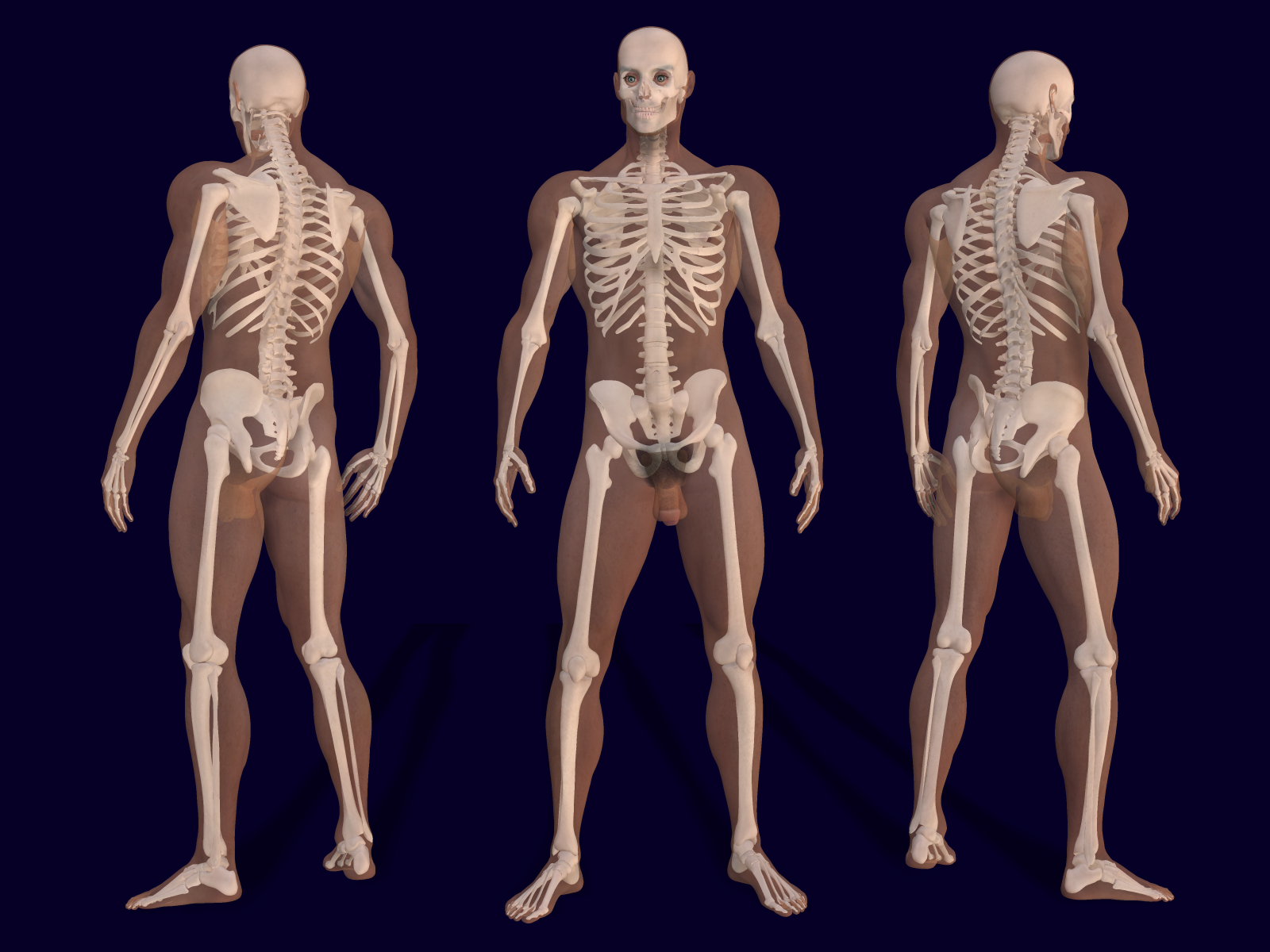
Human skeletal system (female left, male right)
click either image to see expanded views
from Bernhard Ungerer used by permission
Bone organs (gross structure)
Bone number
206 bones is standard / typical (but nearly everyone has more/fewer)
Regions of skeleton
Axial skeleton—forms “axis” of body
- Includes bones of skull, vertebral column, thorax
Appendicular skeleton—forms appendages (arms, legs)
- Includes bones of shoulder/arm/hand and hip/leg/foot
Bone categories
Long bones
- Examples: radius, ulna, tibia, fibula, humerus, femur, metacarpal bones, metatarsal bones
Short bones
- Examples: carpal bones, tarsal bones
Flat bones
- Examples: sternum, ribs, scapula
Irregular bones
- Examples: vertebrae, pelvic bones, skull bones of face
Sesamoid bones
- Example: patella
Long bone structure
Epiphyses (sing. epiphysis) are end regions
- Usually have spongy bone on inside, compact bone on outside
Diaphysis is middle “shaft” region (pl. diaphyses)
- Usually compact bone on outside, cavity on inside
- Medullary cavity contains yellow marrow
- Lined with thin membrane called endosteum
Bone covered with periosteum (dense fibrous sheet) and articular (joint) cartilage
- The dense fibrous connective tissue of the periosteum is continuous with the fibers of bone tissue, as well as with the connective tissue fibers of the deep fascia—thus forming a continuous, strong bond across broad regions of the body.

Long bone structure
Flat bone structure
Short and irregular bones have a similar structure to flat bones
Internal and external table
- Thick sheet of compact bone that forms the hard outer shell of bone
- Covered with periosteum
Diploe (diploë)
- Cancellous (spongy) bone filled with red marrow forming inner region of bone
![]() Review Mini Lesson: Fascial System
Review Mini Lesson: Fascial System
Bone tissue (microscopic structure)
Compact bone
Hard bone forming outer shell of all bone organs
Bone matrix is collagen fibers with apatite mineral (calcium/phosphorus) crystals encrusted on the fibers
- Always starts out as fibrous membrane or cartilage, then turns to bone
- Endochondral ossification — cartilage becomes bone

- Epiphyseal plate is cartilage between epiphysis and diaphysis as they grow together
- Intramembranous ossification — membrane becomes bone
- Fontanels are “soft spots” in infant skull where ossification is not complete
- Allow for deformation of skull during childbirth
- Fontanels are “soft spots” in infant skull where ossification is not complete
- Endochondral ossification — cartilage becomes bone
Osteon (haversian system) is a tapered, cylindrical unit that makes up compact bone tissue ![]()
![]()
- Central canal (haversian or osteonal canal) surrounded by concentric lamellae (layers) of hard bone matrix

- Transverse (Volkmann) canals connect central canals side to side, forming blood networks
- Osteoblasts (“bone makers”) make matrix, then are trapped and now “retired” and called osteocytes (“bone cells”)
- Lacuna (pl. lacunae) are the spaces in which osteocytes are found
- Osteocytes may “come out of retirement” during remodeling
- Canaliculi (“tiny canals”) connect the lacunae (each housing one osteocyte) to each other and to the central canal
- Processes of adjacent osteoblasts are joined by gap junctions, making them a syncytium of cells
- Lamellae (sing. lamella)
- Concentric lamellae form osteons
- Intersititial lamellae, found between osteons, are left over from preliminary osteons and woven bone
- Circumferential lamellae form continuous boundaries at the outer and inner surfaces of the compact bone (around the inner and outer circumference)
Around 21 million osteons in adult skeleton
Each osteon is 100-400 µm in diameter (1 inch = 25,400 µm)
A medium osteon has about 30 lamellae (each about 3 µm)
The central canal is around 50 µm in diameter
Cancellous (spongy) bone
Made up of irregular, fractal trabeculae of hard bone surrounded by red bone marrow (liquid)
- Cancellous bone is also called trabecular bone
Red marrow is myeloid tissue (myelo = “marrow”)
- Hematopoiesis
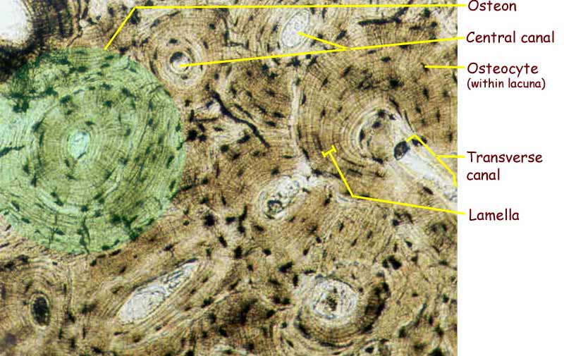
Compact Bone (osteon highlighted in green)
Click image to enlarge it
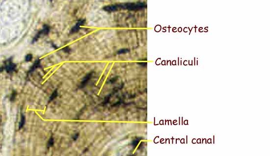
Osteon (closer view)
Click image to enlarge it
Remodeling
Bone is constantly being torn down and built up—this is remodeling ![]()
Role of bone cells ![]()
- Osteoblasts make new bone matrix (using Ca++ from blood)
- Osteoclasts (“bone breakers”) dissolve bone matrix (releasing Ca++ to blood)
Role of hormones
- Calcitonin (CT; from thyroid) increases Ca++ storage (out of blood)
- Parathyroid hormone (PTH) gets Ca++ out of storage (into blood)
Age effects
- More bone is made than is lost until age 25 (usually rapid until puberty, then slows)
- About as much bone is made as is lost 25-50 (can vary)
- More bone is lost than is made 50-120 (usually slight)
- Yellow marrow replaces red marrow, reducing total RBC production
Stress effects
- Mechanical stress can affect bones (fractures, pressure, exercise, etc.)
- Stress increases bone density (therefore, gravity and exercise increase bone density)
- Usually accounts for changes in bone density in old age
Skeletal variations
Sex
- Male — heavier, larger; more defined markings; deep pelvis
- Female — lighter, smaller; less defined markings; shallow/broad pelvis
Age (see section above)
Environment
- Toxins
- Stress (including fractures)—see above
![]() Required—please read the A&P Connect article Skeletal Variations online at the Evolve website
Required—please read the A&P Connect article Skeletal Variations online at the Evolve website
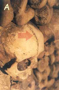
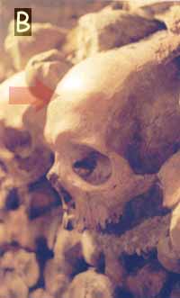
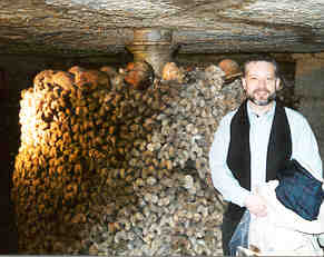 A good place to study normal variations in human skeletons is in the underground ossuary (place for bones) in Paris—also known as the “Paris catacombs.” A good place to study normal variations in human skeletons is in the underground ossuary (place for bones) in Paris—also known as the “Paris catacombs.” 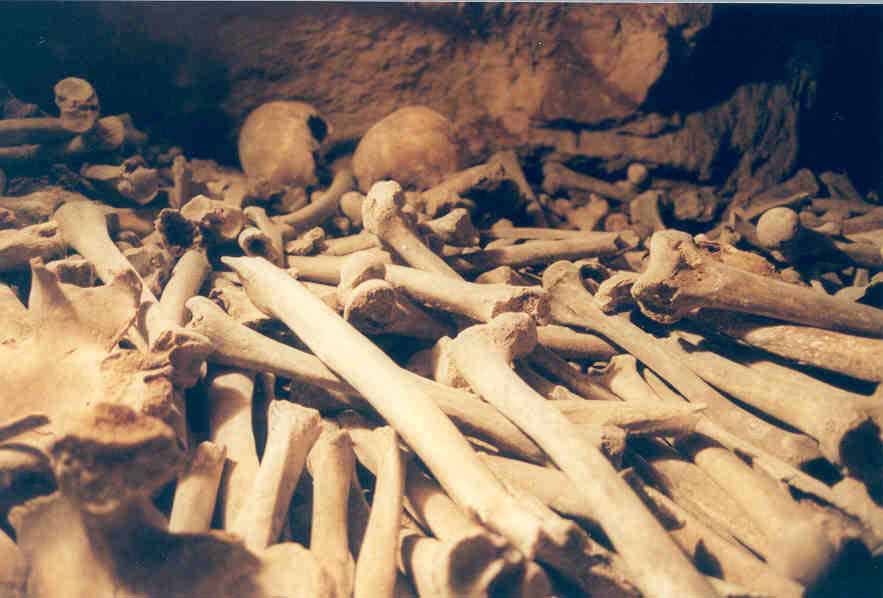 Skeletal remains of thousands of people buried in 18th century cemeteries were moved to abandoned chalk quarries under the streets of Paris and can be visited today. Click each photo to enlarge it. Curious about the Paris catacombs? Click here. Skeletal remains of thousands of people buried in 18th century cemeteries were moved to abandoned chalk quarries under the streets of Paris and can be visited today. Click each photo to enlarge it. Curious about the Paris catacombs? Click here. |
One frontal bone or two?Figure A is a photograph from the Paris catacomb showing a skull with a sagittal suture separating the frontal part of the skull into a left frontal bone and right frontal bone. Figure B is a photograph of a nearby skull that is “standard” in that it has no sagittal suture dividing the frontal bone—it has a single frontal bone. It is very likely that neither individual was aware of these facts while they were alive. This is an example of how the human skeleton can vary from one person to another.
|
Joints
See Tables 9-1, 9-2 and 9-3 in textbook
Definition
Joint is where two or more bones come together (join)
Arthro = joint
- Arthritis = joint inflammation (many causes / types)

Ligaments
- Fibrous structures that connect one bone to another
Structural categories
Fibrous — bones are joined by fibrous tissue
Cartilaginous — bones are joined by cartilage
Synovial — bones are joined at a fluid-filled space lined with synovial membrane
Functional categories
Immovable — bones don’t move relative to one another
- Synarthroses
Slightly movable — bones can move, but not much
- Amphiarthroses
Freely movable — bones have significant movement
- Diarthroses
Fibrous
Fibrous joints are synarthrotic joints
Syndesmoses
- Fibrous bands called ligaments connect the bones
- Example: joints between the distal radius and ulna
Sutures
- Fibrous tissue connects flat bones that fit together like jigsaw puzzle pieces
- Example: joints between flat bones of skull
Gomphoses
- Fibrous tissue connects root of tooth to socket of jaw bone
Cartilaginous
Cartilaginous joints are amphiarthrotic)
Synchondroses
- Hyaline cartilage between bones
- Example: first rib-sternum (costosternal) joint
Symphyses
- Fibrocartilage between bones
- Examples:
- between BODIES of vertebra
- pubic symphysis
- between BODIES of vertebra
Synovial
Synovial joints are diarthrotic ![]()
![]()
Uniaxial — single axis of movement
- Hinge
- Pivot
Biaxial — two axes of movement
- Saddle
- Condyloid
Multiaxial — multiple axes of movement
- Ball and socket
- Gliding
Bursa ![]()
![]()
Types of movements
Angular movements
Angular movements increase or decrease the angle of a joint
Flexion — decreases angle of joint
- Ankle flexion — special case
- Plantar flexion — moves toes inferiorly
- Dorsiflexion — moves toes superiorly
Extension — increases angle of joint
- Hyperextension — goes beyond anatomical position
Abduction — moves part away from midline of body or region
Adduction — moves toward from midline of body or region
Circular movements
Circular movements move body parts in a circle
Rotation — pivots part on its axis
Circumduction — moves distal end of part in a circle-like path
Supination — twisting of limb (e.g. arm or leg) away from median
Pronation — twisting of limb toward median
Gliding movement
Special movements
Ankle movements
- Inversion — move sole toward median
- Eversion — move sole away from median
Forward and back movements
- Protraction — move part anteriorly
- Retraction — move part posteriorly
Up and down movements
- Elevation — move part superiorly
- Depression — move part inferiorly
This is a Learning Outline page.
Did you notice the EXTRA menu bar at the top of each Learning Outline page with extra helps?
Readings, References, & Resources
A&P Core
Betts, J. G., DeSaix, P., Johnson, J. E., Korol, O., Kruse, D. H., Poe, B., Wise, J. A., Womble, M., & Young, K. A. (2013). Anatomy and physiology.
Khan Academy. (n.d.). https://www.khanacademy.org/science/health-and-medicine
Patton, K. T. (2013). Survival Guide for Anatomy & Physiology. Elsevier Health Sciences.
Patton, K. T., Bell, F. B., Thompson, T., & Williamson, P. L. (2022). Anatomy & Physiology with Brief Atlas of the Human Body and Quick Guide to the Language of Science and Medicine. Elsevier Health Sciences.
Patton, K. T., Bell, F. B., Thompson, T., & Williamson, P. L. (2023). The Human Body in Health & Disease. Elsevier Health Sciences.
Patton, K. T., Bell, F. B., Thompson, T., & Williamson, P. L. (2024). Structure & Function of the Body. Elsevier Health Sciences.
Topic Focused
Coming soon!