Learning Outline
Endocrine System
A&P 1
|
Note: you must know the names and sources of all hormones listed in tables of Chapters 18 & 19 (except the hypothalamic releasing hormones). In class, we will focus mainly on the theories of how this system works. The details of individual glands and their hormones will be learned mostly on your own. And (of course) they’ll be coming up as we encounter them later in the course (A&P 2). |
Hormones
Chemical messengers
Released into the blood and having an effect outside the tissue that made it
Source
Endocrine (ductless) glands
- “Regular” endocrine tissue — glandular epithelium
- Neurosecretory tissue — modified neurons
Target concept
Target cells, target tissues, target organs ![]()
Receptors (specific to a particular hormone)
Signal transduction
 Hormones affect only cells that are their targets (that have receptors for that specific hormone). Hormones affect only cells that are their targets (that have receptors for that specific hormone). |
 A hormone will not have an effect on target cells of other hormones. A hormone will not have an effect on target cells of other hormones. |
Pathway
Gland ![]() Blood
Blood ![]() Target tissue (receptor)
Target tissue (receptor)
Two categories 
Steroid — lipids derived from cholesterol
Nonsteroid
- Protein
- Glycoprotein
- Peptide
- Amine (derived from amino acids)
Prostaglandins (PGs)
Lipids (fatty acids)
Have LOCAL effects, but are regulatory chemicals carried in the blood, so they are sometimes called “tissue hormones”
- Paracrine agents or “hormones”
- Substances that regulate other cells in the same tissue
- Autocrine agents or “hormones”
- Substances that regulate the same cell that produced it
- Endocrine hormone
- Substances that regulate cells outside the tissue that produced it
- Classical definition of hormone (one that we’ll use in our course)
Mechanisms of hormone actions
Steroid hormones
Lipid hormones derived from cholesterol
- Examples— testosterone, estrogens (e.g., estradiol), progesterone, aldosterone, cortisone
Mobile receptor model (nuclear receptor model)
- Steroid hormone travels in blood plasma (hormone is temporarily bound to a plasma protein)
- Hormone dissolves through phospholipid bilayer of plasma membrane of target cell
- Hormone binds to receptor inside cell (probably in nucleus), forming hormone-receptor complex
- Hormone-receptor complex triggers the activation of a particular gene (on nuclear DNA)
- mRNA is transcribed at the activated gene
- protein is made using the gene encoded in the new mRNA
- protein causes the hormonal effect
Nonsteroid hormones (peptides, proteins)
Membrane-bound receptors (face out of the target cell) ![]()
- May move into cell if small enough
- Second messenger systems
- cyclic AMP
- Hormone travels in blood plasma
- Hormone binds to its receptor in the plasma membrane
- GPCR (G-protein coupled receptor)
- Hormone-receptor binding activates a G protein (in plasma membrane)
- Activated G protein in turn activates the enzyme adenyl cyclase
- Adenyl cyclase causes ATP to lose two P, becoming cAMP (cyclic AMP [adenosine monophosphate])
- cAMP activates protein kinases (enzymes that activate other proteins/enzymes), producing the hormonal effect
- cyclic AMP
- Ca++-calmodulin
- hormone travels in blood plasma
- hormone binds to its specific receptor in the plasma membrane
- hormone-receptor binding activates G protein, then adenyl cyclase, then protein kinases
- activated protein kinase opens Ca++ channels in plasma membrane
- Ca++ diffuses into target cells (remember, cells HATE calcium, so all the Ca++ was outside the cell to begin with)
- Ca++ binds to calmodulin inside cell
- Ca++-calmodulin complex activates (or inactivates) an enzyme, producing the hormonal effects
- Many drugs are targeted at G-protein-coupled receptors (GPCRs)—about 25% of the top-selling drugs and more than half of all currently used drugs.
Amino acid derivative hormones, such as thyroid hormone, may have intracellular receptors as do steroid hormones
Hormones may work together to regulate target cells
Synergistic effects— combined effect is greater than sum of individual effects
Permissive effects— one hormone allows another hormone to have its full effect
Agonistic effects— two hormones have similar effects
Antagonistic effects— two hormones have opposing effects
Trop(h)ic effects— hormones stimulate development of another gland and secretion of that gland’s hormones
Hormones have diverse functions
Even though our course focuses on one or two main actions for each hormone, each hormone may have hundreds of secondary effects
Important for understanding the complexity of endocrine function
Not all hormones find their targets
Metabolized or excreted
Clinically useful — blood or urine analysis shows hormone levels
Regulation of receptors
Up regulation
- Target cells increase their number of a particular hormone receptor, becoming more sensitive to that hormone
Down regulation
- Target cells decrease their number of a particular hormone receptor, becoming less sensitive to that hormone
Receptor turnover
- Ongoing process of a cell moving receptors to its plasma membrane or nucleus and removing other receptors, all occurring at rates that can be increased or decreased under different conditions
Regulation of secretion
Basal secretion (may be periodic)
- Increase or decrease via feedback controlling mechanisms
- Feedback
- Negative; positive
- Long loop; short loop
- Feedback
Responsive

Cyclic secretion of hormones during female reproductive cycle.
Values plotted from a human study.
Click for a larger image.
Endocrine disorders
|
|
Many tissues in the body act as endocrine glands. Those pictured here are among the glands that have a major endocrine function. |
 |
Hypothalamus-pituitary
Neuroendocrine cells
Neurons that release their transmitters directly into the blood (rather than across a gap to another neuron), so the transmitters then act as hormones
Hypothalamus
Part of brain on inferior side, just in front of (and above) the brain stem
Part of it is nervous tissue, part is neuroendocrine tissue
Pituitary 
Also called hypophysis or hypophysis cerebri
Divided into two parts — anterior pituitary and posterior pituitary ![]()
Anterior pituitary (adenohypophysis)
- “regular” endocrine tissue
- Releasing hormones (and release-inhibiting hormones)from the hypothalamus regulate anterior pituitary secretion
- Hypophyseal portal system
- Major hormones
- Pr (prolactin)
- GH (growth hormone)
- Tropic hormones (trigger development of/secretion by other glands)
- ACTH (adrenocorticotropic hormone)
- TSH (thyroid-stimulating hormone)
- LH (luteinizing hormone)
- FSH (follicle-stimulating hormone)
Posterior pituitary (neurohypophysis)
- Neuroendocrine cells
- Body of cell is in hypothalamus
- Fiber extends from each neuron cell body through the stalklike infundibulum and down into posterior pituitary
- Thus, posterior pituitary hormones are made in the hypothalamus but released from the posterior pituitary
- Oxytocin and ADH (antidiuretic hormone)

Location of hypothalamus and pituitary
Thyroid
Location — below larynx, wrapped around trachea ![]()
Structure
- Two lateral masses and isthmus
- Thyroid follicles
- Follicular cells — release thyroid hormone
- Thyroid colloid — stores thyroid hormone
- Thyroid hormone
- T3 — triiodothyronine
- T4 — tetraiodothyronine
- Follicular cells — release thyroid hormone
- Parafollicular cells
- Calcitonin
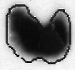 |
Thyroid scan. Dark areas represent uptake of radiactively tagged iodine (I). Iodine is a trace element required for the manufacture of thyroid hormone. |
Parathyroid
Location ![]()
- Imbedded in posterior surfaces of thyroid
Structure
- 4-5 small round bodies made of glandular epithelium
Hormone — parathyroid hormone (PTH)
Adrenal
Also called suprarenal
Location ![]()
![]()
![]()
- On top of each kidney (renal = “kidney”)
Structure ![]()
- Cortex (endocrine)
- Zona glomerulosa
- Mineralocorticoids (aldosterone)
- Zona fasciculata
- Glucocorticoids (cortisone)
- Zona reticularis
- Glucocorticoids (cortisone)
- Gonadocorticoids (testosterone, estrogens)
- Zona glomerulosa
- Medulla (neurosecretory)
- Catecholamines (epinephrine[adrenaline])
Pancreas
Location ![]()
![]()
- Lies within the C of the duodenum, inferior to the stomach
Structure
- Pancreatic islets (of Langerhans) (endocrine)
- Alpha cells (A) — glucagon
- Beta cells (B) — insulin
- Delta cells (D) — somatostatin|
- PP cells (F) — pancreatic polypeptide
- Epsilon (E) — ghrelin
- Acinar cells (exocrine)
Other endocrine glands
Ovaries
Estrogens and progesterone
Testis
Testosterone
Placenta
Human chorionic gonadotropin (HCG)
Estrogens
Progesterone
Placental lactogen
Thymus
![]()
Thymosin
GI (gastrointestinal) mucosa
Several hormones regulate GI function
Heart
Atrial natriuretic hormone (ANH)
Pineal
Melatonin
![]() Please refer to the tables in the Anatomy & Physiology
Please refer to the tables in the Anatomy & Physiology![]() textbook for a summary of these and other additional hormones of the human body.
textbook for a summary of these and other additional hormones of the human body.
Example of homeostatic endocrine regulation
Calcium concentration in blood plasma
- Parathyroid hormone (PTH) increases blood Ca++ by increasing rate of Ca++ breakdown (reabsorption) in bone tissue and by increasing rate of Ca++ in digestive tract
- Calcitonin (CT; from thyroid) decreases blood Ca++ by decreasing rate of reabosorption of calcium in bone tissue
This is a Learning Outline page.
Did you notice the EXTRA menu bar at the top of each Learning Outline page with extra helps?
Last updated: October 22, 2019 at 14:22 pm