Learning Outline
Nervous System 1
Be sure to check these outlines also
Overview of Nervous System
Nervous System Part 2
Neural histology
Two kinds of cells
Neurons = nerve cells
- Transmit / process information
Neuroglia (glia)
- Support
- May also process or modulate information
9:1 ratio of glia to neurons on average in entire nervous system (ratio may be higher in more complex processing centers; estimates vary widely)
Glial cells [neuroglia]
Many types ![]()
- Astrocytes

- Microglia
- Ependyma
- Schwann cells (neurolemmocytes)
- Oligodendrocytes
|
|
Microglia |
Ependyma |


Many functions
- Structural support
- Astrocytes hold neurons/blood vessels in place
- Ependyma line fluid spaces in brain
- Protection
- BBB — blood-brain barrier
- Astrocytes form wall around blood capillaries (small vessels) in brain, keeping chemicals from passing from blood into brain tissue
- Nurturing and development of neurons (e.g., nerve growth factors)
- Housekeeping (clean-up)
- Microglia (small, but can enlarge and become phagocytic cells)
- Electrical insulation of nerves
- Myelin (white lipid in cell membranes) sheath and neurilemma (outer cytoplasm of glial cell) around neuron fibers
- Formed by: Schwann cells (in nerves) and oligodendrocytes (in brain/cord)
- Gaps in myelin sheath are called nodes [of Ranvier]
- Communication
- Chemical signals to each other / to neurons
Overview of neurons and their role in the nervous system
Overview of nervous system
Central nervous system (CNS) is brain and cord
Peripheral nervous system (PNS) is nerves and related structures
Review of nerve reflex arc
Pattern of information/control like a feedback loop
- Receptor (sensitive organ or tip of neuron)
- Sensory neuron (carries info toward CNS)
- Interneuron (may be none or many)
- Motor neuron (carries info away from CNS)
- Effector (usually a muscle or gland)
|
Yikes! Why do we have to know all these details?
|
|
Good question! The following information will seem like overkill to the beginning student. Believe it or not, this is an oversimplification of what we know about how nerve cells work! Even so, the details you learn here will later help you understand essential concepts such as
|
Typical neuron
Excitable cell
Capable of an “impulse” or voltage fluctuation
Types of neurons
Functional categories
- Sensory (afferent) neuron
- Interneuron (association neuron)
- Motor (efferent) neuron
Structural categories
- Multipolar
- Bipolar
- Unipolar
Cell body (soma, perikaryon)
- Conducts impulses toward axon
- Mitochondria replicate here (some move to extensions)
- Rough ER (Nissl bodies), Golgi apparatus
- Manufacture/remanufacture neurotransmitters
- Can be replaced in adulthood (previously thought to be irreplacible)
Cell extensions
Extensions in general are sometimes called nerve fibers or neurites
Dendrites (literally “tree branches”)
- Conduct impulses toward axon
- May have dendritic spines—bumps that connect with other neurons
Axon (just one)
- Conducts impulses away from cell body
- Axon hillock — tapered origin of the axon
- Where impulses add together
- If resulting impuls is of sufficient size, then will travel down the axon
- Where impulses add together
- Axon may be myelinated
- Covered with myelin sheath (neuronal sheath)
- Schwann cells (neurolemmocyte) or oligodendrocytes
- Electrical insulation
- Gaps called nodes [of Ranvier] or myelin sheath gaps
- Allows rapid conduction of impulse from node to node
- Covered with myelin sheath (neuronal sheath)
- Synaptic terminal (axon terminal)
- Store, then release, neurotransmitter molecules
- Varicosities — swellings along an axon
- Function like axon terminals when they link to other cells such as smooth muscle cells
Cytoskeleton
Neurofibrils (intermediate filaments; neurofilaments), microtubules and microfilaments ![]()
Axonal transport system
- Transport system of “railways” and motor molecules that shuttle vesicles and mitochondria from cell body to end of axon and bring vesicles back to cell body
![]() Explore optional animated overviews of nervous system cells
Explore optional animated overviews of nervous system cells

Nerves and tracts
Nerve
Nerve — bundle of nerve fibers in PNS ![]()
Connective tissue component ![]()
- Endoneurium — around individual axons
- Perineurium — around fascicles (bundles of axons)
- Epineurium — around whole nerve

Tract
Tract — bundle of nerve fibers in CNS
- Lack connective tissue component
Gray and white matter
- White matter (white substance)

- Myelinated nerves and tracts (myelin is white)
- Where information is “passing through”
- Gray matter (gray substance)
- Unmyelinated nerves, tracts, cell bodies
- Where information is processed
- Nucleus = area of gray matter in the CNS
- Ganglion = area of gray matter in PNS
|
FYI |
| Gray is standard American English and WILL be used in this course. Grey is standard in other English dialects and will NOT be used in this course. Check out this brief article. |
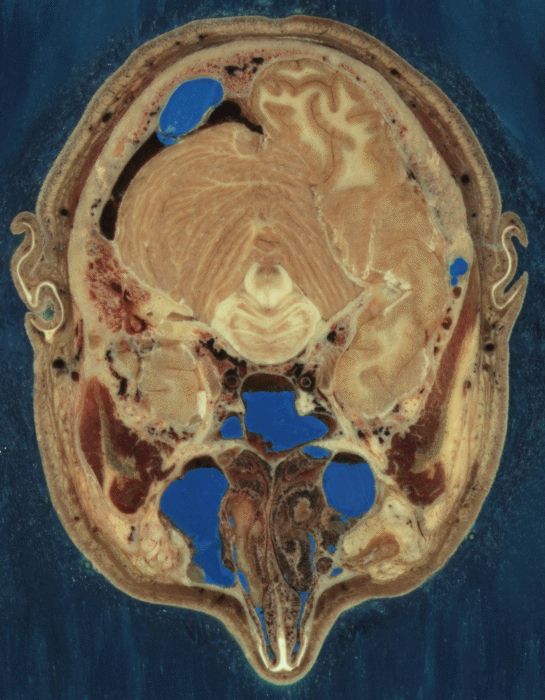 |
These horizontal sections of the human head came from the Visible Human Project and show the distinction between white matter and gray matter in nervous tissue. Click on each image to see a larger, more detailed view |
Nerve impulse
Definitions
Potential = Gradient of electrical potential energy (difference in electrical charge) between two points
Voltmeter = Detects electric potential
- Electric potential is measured in volts (V) or millivolts (mV)
![]() Hint: Print several copies of the figure at
Hint: Print several copies of the figure at ![]() and have them ready in class to take notes
and have them ready in class to take notes
Membrane potential = Electrical gradient maintained across living cell membranes
- Cell voltmeters are arranged so that the sign (- or +) tells the charge on the inside surface of the plasma membrane
Resting membrane potential (RMP) = Membrane potential during rest (in an excitable cell) = -70 mV
Mechanisms
Two ions at play here
Sodium [Na+] and potassium [K+]
- For the sake of our story, these two ions are the ONLY two ions that can move, all other positive ions and all negative ions are impermeant
Na+/K+ pump ![]()
- Also known as Na-K pump or Na+-K+ ATPase, this is a countertransport (antiport) mechanism
- The Na-K pump makes sure that
- Na+ is concentrated outside the cell (remember, cells HATE Na+)
- K+ is concentrated inside the cell (cells LOVE K+)
- In the human body at rest, about one-third of energy used goes toward powering the Na-K pump!
Several gated channels are at play here, too
Stimulus-gated channels are triggered by sensory stimuli or nerve stimuli (neurotransmitters)
Voltage-gated channels are triggered by a fluctuation in voltage (membrane potential)
- The minimum voltage needed to trigger a voltage-gated channel is called the threshold potential (-59 mV)
All of these channels are specific (either allow Na+ or K+ through —not both) ![]()
RMP
There’s more Na+ outside the cell than K+ inside, thus there is an imbalance of too many positive ions on the outside
Produces an RMP of -70 mV
Membrane is polarized—that is, it has a negative pole and positive pole
Local potentials
Fluctuations from RMP caused by activation of stimulus-gated channels in the dendrites/cell body ![]()
- Decremental conduction = they “poop out”
- Local potentials never, ever, ever, ever, ever, reach the end of the axon
- But local potentials are needed to possibly start an action potential, which does reach the end of the axon (more on that later)
- Summation = they can add together
Types of Local Potentials
- Direction of voltage fluction (up or down on the voltmeter)
- Depolarization = if Na+ gates open, Na+ rushes into cell and increases voltage (less negative inside)
- Hyperpolarization = if K+ gates open, K+ rushes out of cell and decreases voltage (more negative inside)
- Triggering stimulus (nerve or sensory)
- Synaptic potential = local potential triggered by chemical signal at a synapse (junction)
- Receptor potential = local potential triggered by a sensory stimulus
Action potentials
If local potential reaches axon, and is a depolarization large enough to reach the threshold potential (-59 mV), then voltage-gated channels in the axon will open —causing an action potential ![]()
- Nondecremental conduction
- Nonsummating (all-or-none events)
Voltage-gated Na+ channels open first, and Na+ rushes in to depolarize the membrane to +30 mV
Then voltage-gated K+ channels open (they’re slow, like your A & P prof, OK?) and K+ rushes out to repolarize the membrane back toward RMP
Peak of +30 mV triggers the next section of axon to open its voltage-gated channels, and the process repeats —and keeps repeating all the way to the terminals at end of axon
- Velocity of conduction
- Continuous conduction
- Action potential moves point-to-point along fiber
- Velocity is proportional to diameter of fiber
- Saltatory conduction — node-to-node conduction by myelinated fibers increases velocity (compared to ordinary point-to-point conduction in nonmyelinated fibers)
- Explore optional animations of regular and saltatory conduction click here
- Myelin disorders
- Disrupt saltatory conduction and thus change speed of nerve signaling, causing coordination problems
- Example: multiple sclerosis (MS)
- Montel Williams (see Climbing Higher
 )
) - Teri Garr (see Speedbumps: Flooring It Through Hollywood )
- Montel Williams (see Climbing Higher
- Explore optional animations of regular and saltatory conduction click here
- Continuous conduction
Action potentials are all-or-none events
Refractory period
- Absolute refractory period — no new action potential can begin
- Relative refractory period — a new action potential can begin ONLY if there is a very large depolarization
- the threshold potential is is very high during this phase
- this means the frequency of action potentials can be higher than normal if the stimulus is very large
Synaptic transmission
Types of synapses
Electrical synapse — gap junctions
Chemical synapse — neurotransmitters & receptors
Parts of a synapse
Presynaptic neuron = the neuron that gets the signal first and is about to transmit it to a second neuron
Synaptic cleft = narrow space separating two adjoining neurons
Postsynaptic neuron = the neuron that gets the signal after having received it from the presynaptic neuron
Mechanism of synaptic transmission
Action potential reaches the presynaptic terminal
High voltage of action potential triggers opening of voltage-gated Ca++ channels in presynaptic terminal
Ca++ flows in and triggers the cytoskeleton to move vesicles containing neurotransmitter to surface and undergo exocytosis (release of contents)
Neurotransmitter diffuses across synaptic cleft
Neurotransmitter molecules bind to receptors in postsynaptic plasma membrane (according to lock-and-key model)
Neurotransmitter-receptor binding causes a change in potential (voltage) in the postsynaptic membrane ![]()
- If it’s depolarization, then it can also be called excitation or facilitation because it gets the postsynaptic cell closer to the threshold potential and therefore closer to the action potential or “excitation”
- If it’s hyperpolarization, then it can also be called inhibition because it inhibits the chances of an action potential in the postsynaptic neuron
Transmission must be “turned off” or it will continue forever ![]()
- Reuptake of neurotransmitter into presynaptic terminal
- May be transported into nearby glial cells (which may release them again for reuptake by presynaptic cells)
- Breaking of neurotransmitter molecule by enzymes
- Loss of some neurotransmitter from synapse by diffusion
Other concepts of synaptic transmission
Direct and indirect signal transduction
- Signal transduction refers to ANY mechanism by which a chemical or other stimulus is interpreted by the cell to cause a change
- Direct mechanisms involve a receptor that is part of the ion channel that responds to the neurotransmitter
- Indirect mechanisms are second-messenger systems that may involve separate receptors that activate G-protein and cAMP to get ion channels to respond
- Many drugs are targeted at G-protein-coupled receptors (GPCRs)—about 25% of the top-selling drugs and more than half of all currently used drugs.
There is always a low-level “baseline” amount of transmitter in every synapse
- That is, you don’t ever really start a synaptic transmission with an empty synaptic cleft
- This is often referred to as “the balance of chemicals” in your brain
- Whether you have sufficient baseline amounts of transmitter in certain neural pathways to allow for normal function of those pathways
- Whether you have sufficient baseline amounts of transmitter in certain neural pathways to allow for normal function of those pathways
- Neurotransmitter baseline can be altered by therapeutic chemicals

- Reuptake inhibitors — block return of transmitter to presynaptic neuron, thus getting baseline amounts in synapse back up to normal
- Enzyme inhibitors — block the breakdown of transmitter molecules at the synapse, increasing the amount of active transmitter present in the synapse
- Reuptake inhibitors — block return of transmitter to presynaptic neuron, thus getting baseline amounts in synapse back up to normal
Postsynaptic cells integrate signals from thousands of presynaptic neurons to determine whether or not the signal will continue along the neural pathway
- Excitatory postsynaptic potential (EPSP) = depolarization in postsynaptic membrane
- Inhibitory postsynaptic potential (IPSP) = hyperpolarization in postsynaptic membrane
- Summation
- Occurs at axon hillock
- Types
- Temporal summation — add up potentials over narrow window of time
- Spatial summation — add up potentials coming from different locations
- Summation is a decision-making process
- Neural pathways can
- Converge (come together)
- Diverge (split apart)
Retrograde signaling — neurons can also send neurtransmitters “backwards” from the postsynaptic cell to the presynaptic cell, further enhancing communication within a neural network
Neurotransmitters and receptors
Receptors
- Can be many different receptors for the same neurotransmitter
Neurotransmitters
- Small molecule transmitters
- Class 1 — Acetylcholine
- Class II — Amines
- Class III — Amino acids
- Class IV — Very small transmitters
- Examples: NO (nitric oxide) and CO (carbon monoxide)
- Large molecule transmitters
- Neuropeptides
- Neurotransmitters can also be classified as “excitatory” or “inhibitory” but the same transmitter may have different effects in different locations
Memory
Proposed mechanisms of memory ALL involve changes at synapses
Learning promotes facilitation of synaptic transmission
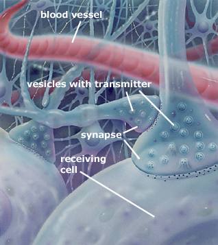 |
This illustration shows several chemical synapses in the nervous system. Notice that a synaptic terminal may have a synapse that influences its output of neurotransmitter. With this arrangement, a neural network can operate a “gateway” in which information can be “cut off” or “enhanced” before moving further along. Source: www.nobel.se |
This is a Learning Outline page.
Did you notice the EXTRA menu bar at the top of each Learning Outline page with extra helps?
Last updated: October 22, 2019 at 14:39 pm