Learning Outline
Nervous System 2
Be sure to check these outlines also:
Overview of Nervous System
Nervous System 1
Overview
Divisions of the nervous system (see Nervous System 1)
Review of tissue types
- White matter — mostly myelinated axons; serves as set of “cables” that transmit information
- Connectome—map of nervous system connections & pathways
- Gray matter — mostly cell bodies and dendrites (many synapses); serves as set of “processing centers” or “control centers”
- Islands of gray matter are called nuclei (CNS) or ganglia (PNS)
Review types of pathways
- Afferent — toward the center
- Efferent — away from the center
| Santiago Ramón y Cajal (1852-1934) is considered by many to be the originator of the modern concept of the nervous system and its organization. Most of this Spanish researcher’s theories about the nervous system and its function are intact today. Although he wanted to be an artist, his father convinced him to follow in his footsteps as an anatomist—a choice that led to a Nobel Prize in Medicine or Physiology in 1906 and many other recognitions. Ramón y Cajal is considered to be a central figure in the history of neuroscience. | 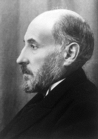 |
Central Nervous System (CNS)
Spinal cord 
Inner gray columns ![]()
![]()
- Anterior, lateral, posterior columns
- Gray commissure (with central canal)
- Integration of spinal reflexes
Outer white columns
- Anterior, lateral, posterior columns (funiculi)
- Spinal cord is mostly white matter
- Ascending tracts and descending tracts conducting sensory and motor information
Features
- Anterior median fissure
- Posterior medial sulcus
- Spinal nerves (31 pairs, all originating on spinal cord)
- All are mixed nerves(sensory & motor fibers) nerves
- Ventral root — motor fibers only
- Dorsal root — sensory fibers only
- Dorsal root ganglion — sensory cell bodies
- Named for segments, as are vertebrae (C1, C2, C3, . . . )
- Cervical (8)
- Thoracic (12)
- Lumbar (5)
- Sacral (5)
- Coccygeal (1)
- All are mixed nerves(sensory & motor fibers) nerves
- Cauda equina (“horse’s tail”)
- Filum terminale (“end string”)
Brain

Located in cranial cavity ![]()
Brain stem
- Three regions — medulla, pons, and midbrain

- Lower level of regulation
- Control centers for respiration, digestive reflexes, cardiac reflexes, etc.
- Eye/ear reflexes
- Corpora quadrigemina (midbrain) (quadri = “four” gemina = “twins”)
- Superior colliculi (eye)
- Inferior colliculi (ear)
- Corpora quadrigemina (midbrain) (quadri = “four” gemina = “twins”)
- Pain modulation
- Pathways for conducting sensory information upward and motor information downward
- Includes much of the reticular activating system (RAS)
- Reticular formation — network of gray matter areas
- “Filters” incoming sensory information and assesses its value/importance
- Stimulates the higher centers (in cerebrum) to stay awake
- Some drugs affect the RAS, for example:
- Barbiturates depress the RAS, reducing alertness
- Amphetamines stimulate the RAS, increasing alertness
- Reticular formation — network of gray matter areas
- Features
- Pyramids (medulla)
- Decussation of pyramids (crossing over of some spinal, left-right & right-left)
- Decussation of pyramids (crossing over of some spinal, left-right & right-left)
- Olives (medulla)
- Corpora quadrigemina (midbrain)
- Cranial nerves
- 12 pairs, 10 originating on brain stem
- Can be primarily sensory, motor, or mixed nerves
- Named by Roman numeral (I, II, III, . . .) beginning with most anterior pair
- Also named descriptively (Olfactory, Optic, . . .)
- 12 pairs, 10 originating on brain stem
- Pyramids (medulla)
 Cerebellum
- Roughly spherical organ attached to posterior side of brainstem
- Wrinkled surface of gray matter
- Regular sulci (grooves) and gyri (raised areas)
- Allows larger surface area and therefore more processing capability
- Cerebellum also has more neurons than all other parts of the brain combined
- Arbor vitae (“tree of life”)
- Interior branches of white matter
- Gray matter gyri are therefore often called called folia (literally “leaves”)
- Connections (“wiring”)
- Interior branches of white matter
- Coordination & planning of skilled muscle activity
- Motor program — set of nervous signals that accomplish a specific set of movements
- Some postural regulation (muscle tone, balance)
- Recent evidence shows it has a much broader role than previously thought, perhaps also being involved “behind the scenes” in many of the cerebrum’s more complex functions.
 Diencephalon
- Literally “the between-brain”
- Just superior to brainstem
- Main regions
- Thalamus
- Oval mass of gray matter
- Two lateral masses joined by thinner intermediate mass
- Acts as a “switchboard” for information heading up to higher centers (in cerebrum)
- Low level of consciousness, sensory perception, emotional response
- Some motor regulation
- Oval mass of gray matter
- Hypothalamus
- Below thalamus
- Mostly small nuclei (areas of gray matter)
- Neuroendocrine link
- Homeostatic control centers
- Center of limbic system
- Limbic system is a ring of structures in different regions of the brain that center around the hypothalamus
- Limbic system allows emotional feelings to occur
- Communicates with conscious centers in cerebrum to allow our thoughts and feelings to be “connected”
- Below thalamus
- Pineal body
- Posterior, cone-shaped structure (pineus = “pine”)
- Another neuroendocrine link
- Acts as part of body’s internal clock
- Has inputs from the eye that help determine time of day, month, year using fluctuations of daylight and moonlight
- Releases the timekeeping hormone melatonin to signal other parts of the body “what time it is”
- Melatonin is an altered form of the neurotransmitter serotonin
- Brain sand
- Calcifications in the pineal make it a good landmark in x-ray and CT imaging
 Thalamus

Cerebrum
- Cortex — gray matter; wrinkled outer surface of cerebrum
- Subdivided into hemispheres and lobes
- Hemispheres — left and right
- Lobes — frontal, parietal, temporal, occipital, insula
- Functional areas — see maps
- Localization — each job is located at a specific place
- Plasticity — some ability to change the place at which a job is done
- Subdivided into hemispheres and lobes

Brodmann areas (BAs) are structural regions of the cerebral cortex.
First identified by Korbinian Brodmann, they are based on the organization of neurons in each regon fo the cortex. These Brodmann areas make up the functional areas outlined on maps in your textbook.
-
- Hemisphericity — concept that one hemisphere is better than the other at certain jobs
- Left — Rt-hand control, spoken & written language, science & math skills, reasoning, sort out parts
- Right — Lt-hand control, music & art awareness, recognizing faces & 3-D objects, insight & imagination, grasps “whole” of parts
- Hemisphericity — concept that one hemisphere is better than the other at certain jobs
![]() Go to the Evolve website and read the required A&P Connect article Specialization of Cerebral Hemispheres
Go to the Evolve website and read the required A&P Connect article Specialization of Cerebral Hemispheres
- Corpus callosum
- White matter band that connects left and right hemisphere, allowing integrated function
- White matter band that connects left and right hemisphere, allowing integrated function
- Association fibers
- White matter/fibers that connect various regions within a hemisphere
- White matter/fibers that connect various regions within a hemisphere
- Basal nuclei (also called cerebral nuclei or basal ganglia)
- Islands of gray matter deep inside cortex
- Coordination of motor activity (motor programming)
- Posture, walking, juggling, and other repetitive motions
- Possible roles in thinking and learning
- Coordination of motor activity (motor programming)
- Functions of cerebrum — consciousness, thinking, problem solving, language, memory, and other complex integrative functions

Coverings and Cavities of the CNS
Coverings of CNS
Bony coverings
- Cranial bones
- Vertebral column
Membranous coverings — meninges (sing. meninx)
- Dura mater (“tough mother”) — fibrous outer covering can stop s (but don’t count on it)
- Falx cerebri (falx = “sickle”)
- Dural sinuses
- Falx cerebelli
- Tentorium cerebelli
- Falx cerebri (falx = “sickle”)
- Arachnoid mater (“spidery mother”) — wispy middle covering
- Subarachnoid space — filled with cerebrospinal fluid (CSF)
- Pia mater (“delicate mother”) — super-thin coating of brain and cord; innermost meninx

Fluid spaces
Cerebrospinal fluid (CSF)
- Filtrate from blood of choroid plexus
- Functions
- Support/cushioning
- Fluid for sampling/analysis
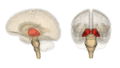
Ventricles and canals ![]()
![]()
![]()
- Lateral ventricles (left and right)
- Separated by septum pellucidum
- Third ventricle
- Cerebral aqueduct (of Sylvius)
- Fourth ventricle
- Lateral & median foramina
- Central canal
- Subarachnoid space
- Reabsorption of CSF
- Reabsorption of CSF
![]() Go to the Evolve website and read the required A&P Connect article Hydrocephalus
Go to the Evolve website and read the required A&P Connect article Hydrocephalus
Measuring brain activity
Electroencephalogram (EEG)
Measured in waves/sec or Hertz (Hz)
Show relative level of activity of a whole brain region
- Alpha waves (8-13 Hz) — waking, relaxed
- Beta waves (13+ Hz) — waking, attentive
- Theta waves (4-7 Hz) — drowsy
- Delta waves (<4 Hz) — deep sleep
 EEG wave.
This is a one-second interval . . . what category does it belong to? Are the waves chaotic or periodic?
Sleep
Two states
- REM (rapid eye movement) — dream state
- SWS (slow wave sleep) — deep, dreamless sleep
Swing back and forth between states
- 90-minute cycles
- Deepest at beginning of sleep period; gets less deep toward morning
![]() Go to the Evolve website and read the required A&P Connect article Sleep Stages
Go to the Evolve website and read the required A&P Connect article Sleep Stages
PET
Positron Emission Tomography
- Nuclear materials emit “positrons” that are detected by sensors
- Tomography is “making pictures of sections (cuts)”
Can be used to visualize functions (for example, using oxygen or glucose at different rates in different parts of the brain)
 |
PET scan showing the more active areas in red and the least active areas in blue. This image shows a horizontal section of the cerebrum. |
![]() Go to the Evolve website and read the required A&P Connect article Brain Studies
Go to the Evolve website and read the required A&P Connect article Brain Studies
Peripheral nervous system (PNS)
Cranial nerves (12 pairs, 10 originating from brainstem)
- Plus cranial nerve 0 (terminal nerve)
Spinal nerves (31 pairs originating from spinal cord)
Both cranial and spinal nerves have numerous branches
- Plexus (literally “braid”) is network formed by intermingling of fibers from several different spinal nerves
![]() Go to the Evolve website and read the required A&P Connect article Nerve Zero
Go to the Evolve website and read the required A&P Connect article Nerve Zero
Somatic nervous system (SNS)
Sensory — conscious sensation (vision, muscle position, touch, etc.)
- General principles of sensory reception
- Sensory receptors — structures that are sensitive to specific changes in their surrounding environment (either internal environment or external environment, depending on their location in the body)
- Receptor potential — voltage fluctuation that occurs in response to a change (sensory stimulus); it may or may not generate an action potential, depending on whether it (in combination with other receptor potentials) reaches the threshold potential at the axon
- Sensory adaptation — gradual cessation of response to a specific stimulus; in short, many receptors “fatigue” after the initial onset of a stimulus and stop responding to it
- Dermatomes — sections of the skin that correspond to pathways taken by sensory receptors
- Types of sensory receptors
- Functional categories (based on modality — what the receptor is sensitive to)
- Mechanoreceptors — sensitive to mechanical stimuli such as pressure, vibration, stretch, tilt, etc.
- Thermoreceptors — sensitive to changes in temperature (within a specific range)
- Photoreceptors — sensitive to changes in the intensity and color of light
- Chemoreceptors — sensitive to changes in chemistry (presence or absence of certain chemicals)
- Pain receptors — sensitive to damage or stimuli that may signal potential damage to tissues; any receptor can act as a pain receptor if overstimulated
- Structural categories (based on where in the body the receptor is located)
- Exteroceptors — near the exterior surface of the body (skin/mucous membranes)
- Interoceptors — located within internal structures of the body (muscles/organs)
- Functional categories (based on modality — what the receptor is sensitive to)
- General senses
- Touch
- Pressure
- Temperature
- Proprioception (muscle sense)
- Pain
- Special senses
- Vision — intensity and color of light formed into an image
- Hearing — intensity (loudness) and pitch of sounds
- Smell (olfaction) — intensity and type of aromatic molecules present in air
- Taste — intensity and category (sweet, sour, salty, bitter, umami) present in solution in mouth/throat
- Static equilibrium — body position relative to gravity
- Dynamic equilibrium — change in direction or speed of body movement
- Senses . . . they’re all in your head!
- What you see (or hear, smell, etc.) isn’t all the information in your environment
- Information that is detected, doesn’t necessarily make it to your consciousness
- Information that does enter consciousness has been changed
Motor — conscious (voluntary) control of effectors (skeletal muscles)
- Only one neuron outside the CNS
- Cholinergic — releases the neurotransmitter acetylcholine (ACh)
- Motor programs generated at any of several levels:
- Spinal cord
- Cerebellum
- Cerebral nuclei
- Motor cortex
- Interaction among levels
- Somatic motor tracts
- Pyramidal tracts go through the pyramids of the medulla
- Direct to effectors — distal skeletal muscles
- Fine, skilled movements
- Extrapyramidal tracts do not go through the pyramids
- By way of brainstem nuclei to effectors — proximal skeletal muscles
- Posture; gross movements
- Pyramidal tracts go through the pyramids of the medulla
Autonomic nervous system (ANS)
Sensory — subconscious sensation (oxygen levels, glucose levels, blood pressure, etc.)
Motor — subconscious (involuntary) control of effectors (smooth & cardiac muscle, glands, adipose, and others)
- Sympathetic division — stress (“fight-or-flight” response) reaction
- Primes body for intense skeletal muscle activity to resist (fight) or avoid (flight) a stressor (anything perceived to be a threat to homeostasis)
- Parasympathetic division — maintenance, rest-and-repair
- Counterbalances functions of sympathetic division
- Dual innervation — when effectors are “hooked up to” both divisions at once (that is, a dually innervated effector is a muscle or gland that receives signals from both the sympathetic and parasympathetic divisions)
- Sometimes the effects are antagonistic
- Example: sympathetic speeds up the heart and parasympathetic slows down the heart
- Sometimes the effects are cooperative
- Example: in sexual response, parasympathetic is responsible for initial excitement; at orgasm, sudden switch to sympathetic signals leading to resolution [reversal] of changes that occurred during excitement)
- At dually innervated autonomic effectors
- Sometimes the effects are antagonistic
- Dual innervation requires special transmission mechanisms
- Two neurons between CNS and effector (compare to somatic motor pathway — one)
- Preganglionic neuron and postganglionic neuron
- Autonomic ganglion is where the neurons synapse
- Sympathetic division — preganglionics are generally shorter
- Sympathetic chain ganglia
- Sympathetic chain ganglia
- Parasympathetic division — preganglionics are generally longer
- Cholinergic fibers
- Release acetylcholine (Ach)
- All preganglionic fibers
- All parasympathetic postganglionic fibers
- Sympathetic postganglionic fibers (only those to singly innervated effectors)
- Adrenergic fibers
- Release norepinephrine (NE)
- Also known as noradrenaline (NA)
- Sympathetic postganglionic fibers (dually innervated effectors)
- Thus, at dually innervated effectors there are two different neurotransmitters
- Release norepinephrine (NE)
- Different classes of receptors allow for different effects in different pathways
- Cholinergic receptors
- Nicotinic (N)
- Muscarinic (M)
- Adrenergic receptors
- Alpha (a)
- Beta (ß)
- There are many subtypes for each class
- For example, a1 and a2 (and further sublevels such as a1a and a1b, or further to a1a1 and a1a2)
- Knowing subtypes permits us to understand different effects in different tissues
- Also allows us to develop drug therapies that target specific autonomic effectors (and not all of them)
- Cholinergic receptors
- Cotransmission
- Nonadrenergic-noncholinergic (NANC) transmission
- Additional transmitters are released, increasing the possible complexity of signaling
- Stress response
- Group of adrenergic responses that promote muscle function, for example:
- Increased HR, cardiac output
- Increased respiration rate, volume
- Constriction of vessels in viscera, skin
- Increased sweating
- Glycogenolysis in liver
- Lipolysis in adipose tissue
- Decreased immune response
- Regulates skeletal muscle ion pumps to reduce fatigue
- Group of adrenergic responses that promote muscle function, for example:
 |
Claudius Galen first proposed the term “sympathetic nerves” in the first century CE. He believed that these nerves carried “the sympathies” because they are involved in visceral emotional reactions, as when “the heart leaps with joy” or “aches with loneliness.” |
This is a Learning Outline page.
Did you notice the EXTRA menu bar at the top of each Learning Outline page with extra helps?
Last updated: October 22, 2019 at 14:26 pm